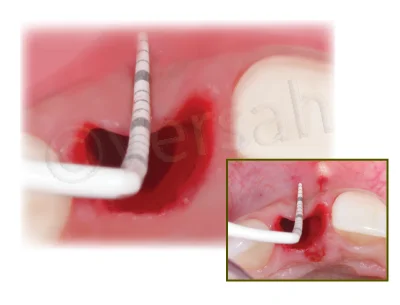
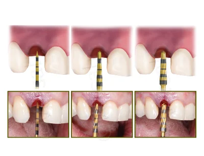
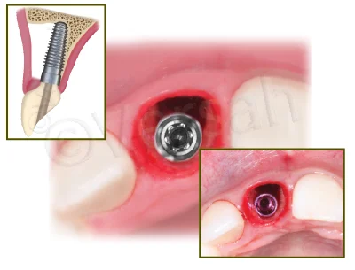
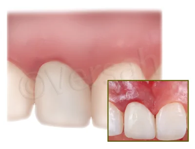
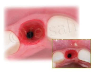
Immediate Dentoalveolar Restoration (IDR) II Technique

Overview

Indicated in cases with total loss of buccal wall in combination with thin periodontal biotype or gingival recession. IDR II Protocol will explain how
to manage compromised sockets with low or no remaining bone in combination with immediate implant placement, bone reconstruction and
provisional fabrication in a single procedure using a combination of bone and soft tissue graft harvested from maxillary tu berosity.
• EXTRACT DAMAGED TOOTH AND REMOVE GRANULATION TISSUE AND REMAINS OF PERIODONTAL TISSUE.
• USE THE DENSAH@ BURS IN OD MODE (CCW ROTATION) TO PREPARE THE IMPLANT SITE AND DENSIFY THE BONE BED.
• ANCHOR THE IMPLANT INTO THE PALATAL WALL.
• HARVEST THE TRIPLE GRAFT AND FILL THE GAP.
• PLACE A MANUFACTURED SCREW RETAINED PROVISIONAL RESTORATION WITH AN ADEQUATE EMERGENCE PROFILE (CEMENTED RESTORATION
MUST NOT BE UTILIZED).
• ALLOW A MINIMUM 4 MONTHS HEALING ANDPLACE A SCREW RETAINED FINAL PORCELAIN CROWN

Step 1:

Extract the damaged tooth using a minimally invasive procedure to
preserve the remaining bone. Completely remove the granulation tissue
and the remains of periodontal tissue or any granulation tissue by
carefully usi ng the curettage technique in the socket. Total loss of the
buccal wall might be common and evident.

Step 2:

Use the Densah@ Burs to prepare the implant site. Start with the
Densah@ pilot drill, in clockwise mode, to a depth related to the planned
implant length. Depending upon the implant type and diameter, follow
with wider Densah@ Burs corresponding with the Implant System
Drilling Protocol.* Starting with the smallest Densah@ Bur, run the
Densah@ Burs in OD mode (counterclockwise, with speed 1000 rpm with
copious irrigation).

Step 3:

Anchor the implant into the remaining palatal bone in the 3-D position.

Step 4:

Harvest the triple graft (connective tissue, cortical and cancellous bone
in one single piece) from the maxillary tuberosity. Insert the triple graft
into the buccal defect aspect of the receptor site to recover hard and
soft tissue that is lost.

Step 5:

Compact particulate autogenous bone harvested from the maxillary
tuberosity to fully fill the gap around the implant, especially in the
buccal aspect.

Step 6:

Place a manufactured screw retained provisional restoration with an
adequate emergence profile.

Step 7:

Allow a minimum 4 months of healing for soft tissue to become stable
in volume and have adequate thickness. A screw retained final
restoration is recommended. Maintenance of the anatomical contour of
soft tissue can be observed.